|
|
|
|
|
|
 |
|
|
|
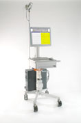
SACS® Video EEG Workstation is the ultimate solution for long-term monitoring. It gives an optimal performance in both networked solutions and as a stand-alone system. All SACS® workstation products are built on the user friendly, safe and modular SACS® platform. With some modules differing the SACS® Video EEG Workstation basically is the same system as the SACS® EEG Workstation, a video input module and a video display module are added and the layout and parts of the user interface are adapted to the needs in long term monitoring. The EEG signals and the digital video signals are both stored in a local SACS® database or in the powerful SACS® Server Database. The video EEG can be reviewed on any SACS® Review Workstation.
MPEG video subsystem
The digital video signal is MPEG coded. Good image quality and high data compression are obtained by the MPEG coding. The user can set the image quality, i.e. the MPEG bite rate. The camera can automatically turn into IR mode when it is dark and an optional IR-light can be applied for night recordings.
Perfect synchronization between Video and EEG
The synchronization between video and the EEG signal is one video frame. The video frame position is shown as a vertical bar on the EEG. If this bar is grabbed by the mouse cursor and moved along the EEG the video is constantly updated and played. The video can be reviewed in slow or fast motion, from 0.25 to 4 * real speed. On the SACS® Video EEG Workstation the EEG and the video in the internal buffer can be reviewed during recording.
Internal Buffering – Review during recording
An internal buffer in the system allows EEG and video for several hours to be reviewed during the recording. The buffer time is hardware dependent. EEG and video are represented in the user interface module by a timeline. Marked events, sections with permanently stored EEG and video as well as the current positions of the EEG and video windows are displayed on the time line. The time line has zoom and tooltips. Mouse clicking on the time line can position the EEG and video windows. Paging can efficiently be made by multiple modes. Review and editing during recording can easily be made in a separate EEG window.
Permanent storage
Data from the internal buffer can by multiple modes be selected for permanent storage. There is no need for storing data continuously; interesting part can be reviewed and stored within the buffer time. If a section of the recording is permanently stored or not is clearly shown by multiple indicators.
Technical Data
Preamplifier:
Inputs 34/64 inputs (27 EEG, 6 polygraphy, 1 respiration )
Input impedance:
100MOhm
Input noise:
< 1,5 microvolt
Common mode rejection ratio:
>100 dB at 50 Hz
Electrode impedance measuring:
LED display, limit values
selectable 5-10-20-50 kOhm
Signal transfer:
Digital via optical fiber cable, electrical separation from the instrument.
Cable:
Standard 5m, maximum 25m without additional amplifier
Sampling frequency:
125 - 1000 Hz
Resolution:
16 bit
Bedside pushbutton:
Yes
Video and audio
Video input:
Composite or s-video
Video standard:
PAL or NTSC
Coding:
MPEG-1
Frame rate:
PAL: 25 frames/s
NTSC 29.97 frames/s
Frame size:
PAL 352x288
NTSC 320x240
Video bit rate:
192 – 4000 kbits/s (>1200 recommended)
Audio sampling:
32 - 44.1 - 48 kHz
Audio bit rate:
32 – 384 kbits/s
Computer
Processor:
Pentium III 733 MHz standard,
needs > Pentium 133 MHz
Harddisk 9 Gbyte or more
Memory 128 Mbyte
Graphics Matrox G400, 16Mbyte
Monitor 21” CRT w. resolution 1600x1280 or 18.1” flat panel w. resolution 1280x1024
Archive
SACS® Server database or CD/DVD-RW |
|
|
|
|
|
|
|
 |
|
|
|
Impedance measurement
Electrode impedance can be measured on demand or scheduled. The display can be set up to warn for to high impedance. When using intracranial electrodes the impedance measurement is inactivated.
Event marks
SACS® Workstation offers five main types of event that the user can add to the recording.
1. Momentary events e.g. swallow.
2. Flexible user defined momentary events. The user defines the event, which can be used for the current recording.
3. States, e.g. wakefulness, status of the eyes and head position. The states are grouped and the values in each group neutralize each other. The states for the current EEG window can be displayed.
4. Free text notations.
5. Clips, for use as bookmarks.
Events can be added, moved and removed as long as a recording or a review session is open. When a recording or a review session is closed no event can be changed to ensure medico legal safety. All events and changes can be traced.
Montages
There can be an unlimited number of user-defined montages. In common average montage a powerful tool for artifact rejection can be used.
Data storage in a powerful database solution
The database solution is highly scalable to fit all needs from stand-alone system to networked solutions serving large hospitals, regions or commercial health care organizations. All data, the EEG as well as the video signal, are compressed and stored within the database, which makes storage and communication safer and access faster and more reliable compared to EEG stored in files administrated by databases. The SACS® Server Database is a true server database, which offers the ultimate performance needed in digital Video-EEG.
Data can be reviewed and edited on one or many remote SACS® Review Workstation during the recording.
The permanently stored sections of an ongoing Video EEG recording can safely be reviewed and edited. Sections to be archived are easily marked and the ongoing recording can be reviewed ‘as reduced’. There is no need for frequent restarting of an ongoing recording. Data reduction of very large recordings over several days is performing very fast.
Database interface supports workflow.
The database interface works like the Windows Explorer interface. The tree structure can be set up to reflect the workflow at the laboratory and it is possible to move recordings by drag and drop technique. The database interface can also be run as a stand-alone program.
Signal measurement cursor
Measurement cursor in EEG windows gives numbers on actual time, time intervals, amplitudes, and frequencies.
Multiple display windows
There are no limitations in how many display windows that can be used. Different montage and filter setting can be shown in different windows. All windows can be synchronized.
Export and Import
Export and import can be customized to any defined file format.
|
|
|